Spatial and
Single-Cell
Proteomics
Spatial and
Single-Cell
Proteomics
Analyzing protein patterns
in single cells with spatial
coordinates
Analyzing protein patterns
in single cells with spatial
coordinates
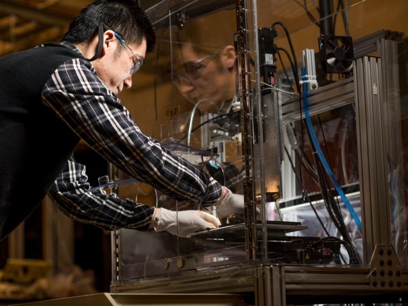
To enable proteomic studies on single cells, PNNL researchers developed the Nanodroplet Processing in One Pot for Trace Samples, or nanoPOTS, platform. This platform for nanoscale proteomics enhances the efficiency and recovery of sample processing and minimizes surface losses by downscaling processing volumes to less than 200 nL. When combined with ultrasensitive liquid chromatography-mass spectrometry analysis, the nanoPOTS platform provides in-depth proteomics analyses of biological samples as small as single cells.
(Photo by Andrea Starr | Pacific Northwest National Laboratory)
Organs and tissues contain many different cell types and subtypes—each with a unique function mediated mainly by proteins and their post translational modifications. Researchers use proteomic measurements to analyze an entire set of cellular proteins expressed at a given time. Most methods for these measurements require samples containing millions of cells, which means that information about localized function is lost.
To enable proteomic studies on single cells, PNNL researchers developed the Nanodroplet Processing in One Pot for Trace Samples, or nanoPOTS, platform. NanoPOTS enhances the efficiency and recovery of sample processing and minimizes surface losses by downscaling processing volumes to less than 200 nL.
When combined with ultrasensitive liquid chromatography-mass spectrometry analysis, the nanoPOTS platform provides in-depth proteomics analyses of biological samples as small as single cells.
We are making many of these first-of-their kind measurements to study unique features of cell populations, including functions, differentiation, and rare subpopulations of cells. Our researchers have tracked the development of sensory hair cells from the inner ear at single cell resolution, identified protein markers of different cell populations from individual human lung cells, profiled proteins in single circulating tumor cells pulled from blood samples, and looked at proteins from pancreatic cells involved in the development of type I diabetes. We can identify protein post-translational modifications in samples with less than 800 cells.
Using spatial proteomics to image tissue proteomes
The location of different cell types in a biological tissue varies in complex ways that direct tissue function. However, the bulk extraction processes currently needed to capture enough protein for proteomic analysis blur the crucial spatial information that helps scientists understand how tissues work together for body functions.
We are combining the sensitivity of the nanoPOTS platform with laser capture microdissection to create an automated method for imaging tissue proteomes. This approach allows us to study tissue substructures and cellular microenvironments at a high spatial resolution not possible before.
Precise and localized analysis of proteins in tissue revealed interconnected molecular networks involved in lung development. We identified protein signatures of a heart attack in fluid pulled from tiny tissues in the kidneys.
Click to view our team of experts who focus on proteomics research in the Biology Division and the Environmental Molecular Sciences Division.
