November 12, 2026
Feature

Researchers Create First Complete 4D Model of a Sun-Fueled Microbe

Molecular biology, genomics, and imaging reveal cellular controls that could inform future biotechnology experiments

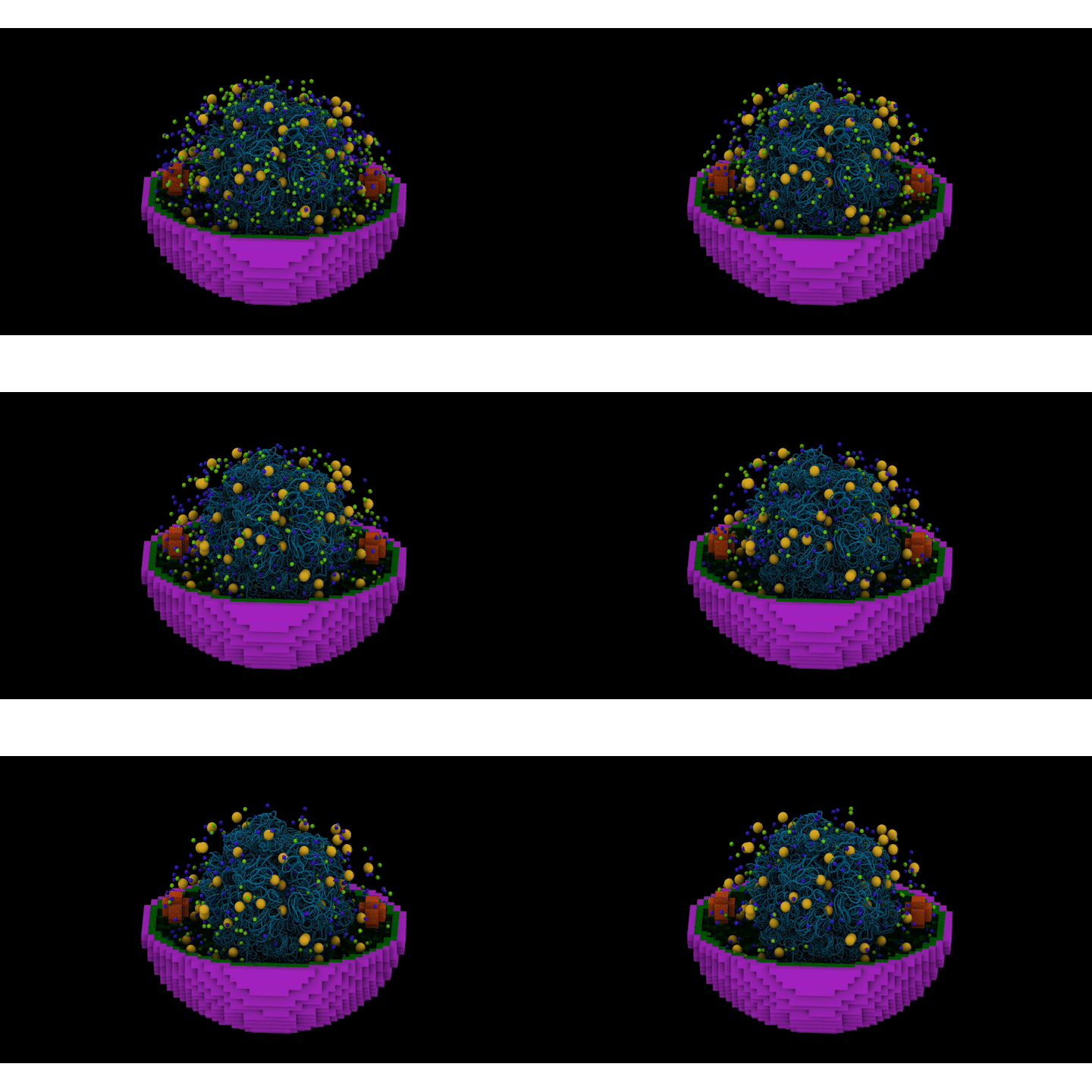
Visual representation of a photosynthetic microbe displaying detail down to the molecular level. The bottom part of the cell is shown in purple and other intricate details are shown in blue, green, yellow, and red. The cell is displayed on a black background.

A four-dimensional whole-cell model shows the locations of key structures and proteins inside a photosynthetic microbe at dusk, helping researchers understand how the cell responds as light changes.

(Image created by Aaron Chan | University of Illinois at Urbana-Champaign)

For some tiny microbes, access to light is essential for life. Organisms that convert light and carbon dioxide to the energy they need to live must continually adjust when light conditions change. Understanding how they maintain stable activity over time could support efforts to use microbes to produce fuels, chemicals, materials, and other useful products.

Now, a research team led by scientists at Pacific Northwest National Laboratory (PNNL) and the University of Illinois Urbana-Champaign have made huge strides in understanding single-celled photosynthetic bacteria, which can convert sunlight to energy like a plant does. They recently published the first four-dimensional whole-cell model of a photosynthetic bacterial cell in the Biophysical Journal.

The model connects the microbe’s three-dimensional structure with changes in gene activity, proteins, and metabolism over time. It could eventually help scientists develop computer simulations, plan laboratory experiments, and reduce some of the trial and error involved in developing microbes for biotechnology.

“This study gives us a detailed view of how an entire photosynthetic cell responds when its energy source changes,” said Margaret Cheung, a computational scientist at PNNL and a corresponding author of the study. “For future biotechnology applications, researchers need to understand not only how microbes produce useful compounds, but also how they maintain stable activity when conditions fluctuate.”

A 4D rendering of a photosynthetic microbe displayed in bright colors (purple, yellow, green, blue, and red) and displayed on a black background.
A four-dimensional whole-cell model follows changing molecular activity inside a photosynthetic microbe, helping researchers understand how the cell maintains stable functions as light conditions change and informing future biotechnology research. (Visual created by Aaron Chan | University of Illinois Urbana-Champaign)

Building a virtual photosynthetic microbe

The team studied Prochlorococcus marinus MED4, a tiny bacterium that uses sunlight for energy and provides a model for studying the essential processes of photosynthetic cells.

The researchers created a detailed digital representation of an entire MED4 cell. The model maps important structures and molecules in three dimensions and follows how they move and interact over time, providing a four-dimensional view of the cell.


Computational scientist Margaret Cheung holds a laptop computer while standing in front of computer stacks.
Margaret Cheung, a computational scientist at Pacific Northwest National Laboratory, led a team in partnership with the University of Illinois Urbana-Champaign that created the first four-dimensional whole-cell model of a photosynthetic bacterial cell. (Photo by Andrea Starr | Pacific Northwest National Laboratory)

To determine where key structures are located, researchers used cryo-electron tomography, an imaging technique that produces detailed three-dimensional views of frozen cells. The images allowed the team to map internal structures involved in capturing light, producing proteins, and carrying out carbon fixation.

The team also collected coordinated time-series measurements of gene activity and proteins taken at matching times as light conditions changed. Researchers studied MED4 cultures as they shifted from darkness to light and from light to darkness. They also examined cultures that remained in darkness or light, helping them distinguish immediate responses to light from changes controlled by the cell’s internal clock.

The researchers combined the imaging and time-series measurements with physics-based computer simulations to create the whole-cell model. Large-scale computing resources supported simulations of molecular activity throughout the cell.

Collecting these different types of information together allowed the team to connect changes occurring across the cell instead of examining each type of biological information separately. The model and its supporting dataset provide resources that scientists can expand, use to test new questions, and adapt to study other photosynthetic microbes.

“Whole-cell models depend on experimental measurements that show what a cell is doing over time,” said Zan Luthey-Schulten, a University of Illinois Urbana-Champaign chemistry professor and corresponding author of the study. “These coordinated measurements allowed us to test the model as light conditions changed and uncover cellular controls that would be difficult to observe through experiments alone.”

Finding the cell’s light-sensitive switch

The research also revealed an important role for CP12, a small protein that helps photosynthetic organisms regulate carbon fixation—the process cells use to turn carbon dioxide into carbon-based molecules needed for growth.

In darkness, CP12 joins with two carbon-fixation enzymes to form the “dark complex,” named because it assembles when light is unavailable. The complex holds the enzymes in an inactive state, preventing the cell from carrying out parts of carbon fixation when it cannot obtain energy from light.

When light returns, chemical changes to CP12 and the enzymes cause the complex to break apart. This releases the enzymes so the cell can resume producing molecules needed for growth.

Because this switch acts directly on existing enzymes, it can adjust carbon fixation more quickly than processes that require the cell to change gene activity and produce new proteins. The model also showed that the locations of proteins and structures within the cell help reduce the effects of random variations in molecular activity. Together, these controls help the microbe respond to changing light while maintaining stable cellular functions.

Taken together, the model offers a framework for investigating how photosynthetic cells balance energy use, growth, and stability. That understanding could inform future research in biotechnology, synthetic biology, and biomanufacturing.

This research is supported by the Biopreparedness Research Virtual Environment (BRaVE), a Department of Energy (DOE), Office of Science, Biological and Environmental Research program. A portion of this research was performed on a project award from the Environmental Molecular Sciences Laboratory (EMSL), a DOE Office of Science User Facility sponsored by the Biological and Environmental Research program and located at PNNL. Computational allocations were supported by a 2024 Advanced Scientific Computing Research Leadership Computing Challenge award and EMSL’s Tahoma high-performance computing facilities.

###

About PNNL

Pacific Northwest National Laboratory draws on its distinguishing strengths in chemistry, Earth sciences, biology and data science to advance scientific knowledge and address challenges in energy resiliency and national security. Founded in 1965, PNNL is operated by Battelle and supported by the Office of Science of the U.S. Department of Energy. The Office of Science is the single largest supporter of basic research in the physical sciences in the United States and is working to address some of the most pressing challenges of our time. For more information, visit the DOE Office of Science website. For more information on PNNL, visit PNNL's News Center. Follow us on LinkedIn, Facebook, YouTube, Instagram and X.

Published: November 12, 2026