PNNL has developed a next-generation electrical resistivity tomography system for DOE that uses E4D software and AI-enhanced modeling to produce real-time subsurface images that help guide environmental remediation decisions.
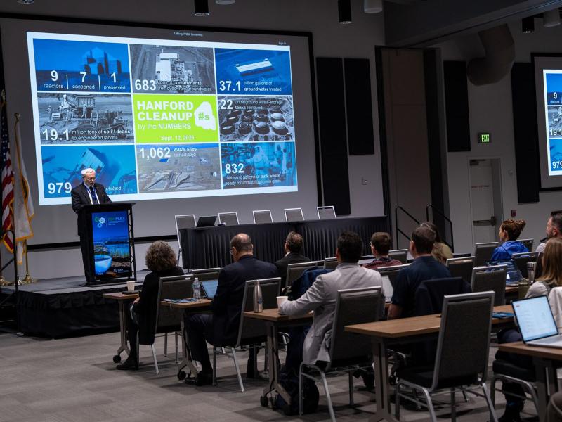
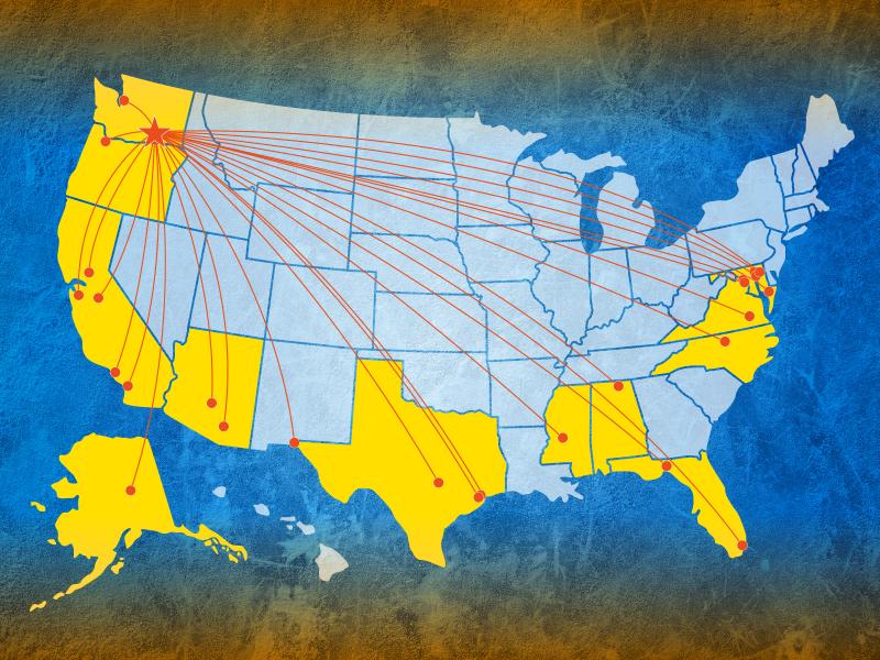
RemPlex 2025 Global Summit on Environmental Remediation attendees share knowledge about cleanup and monitoring of complex sites worldwide; more than 100 presentations are posted online.
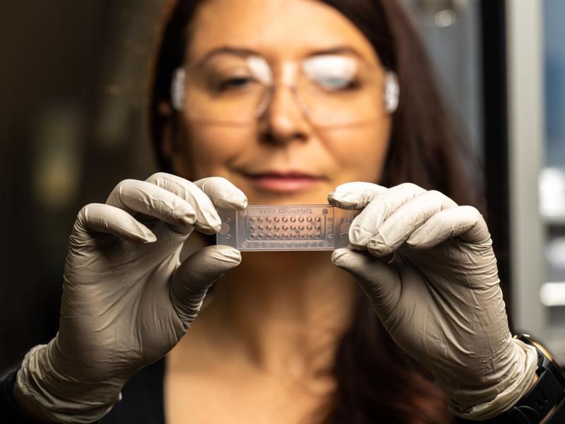
Researchers developed a robust, cost-effective, and easy-to-use cap-based technique for spatial proteome mapping, addressing the lack of accessible proteomics technologies for studying tissue heterogeneity and microenvironments.
This project sought to assure that research activities centered around different sampling and monitoring efforts in northwest Ohio would not disturb any historical cultural resources.
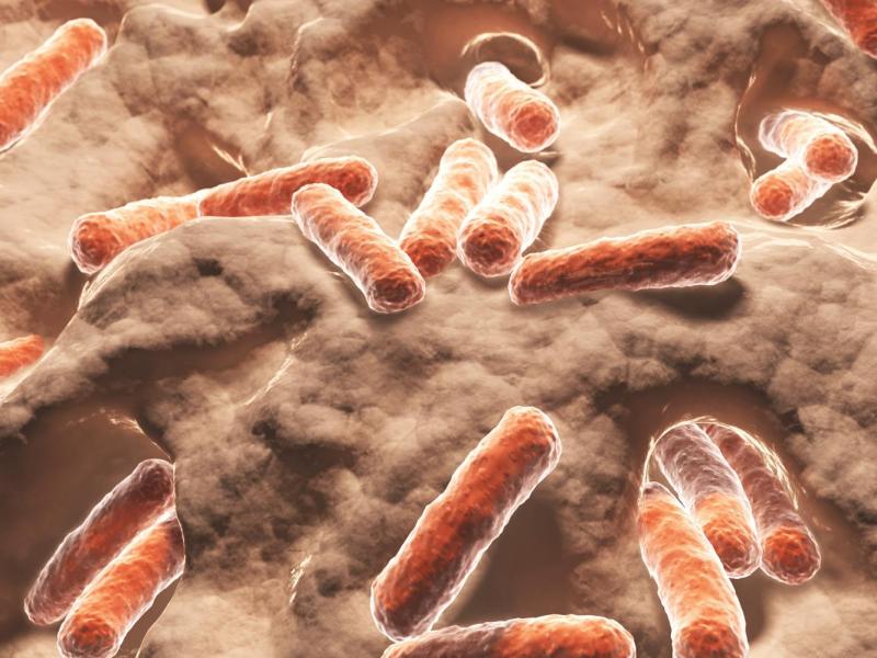
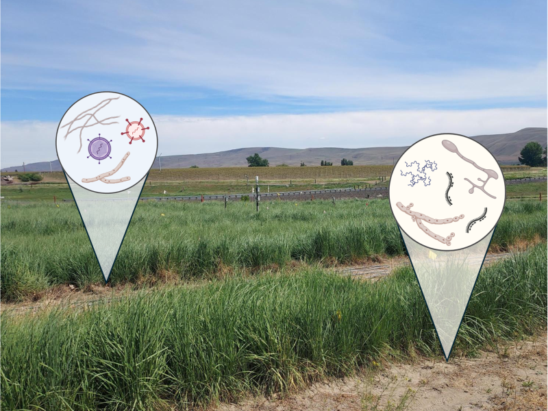
Despite the widespread presence of RNA viruses in soils, little is known about the relative contributions and interactions of biological and environmental factors shaping the composition of soil RNA viral communities.
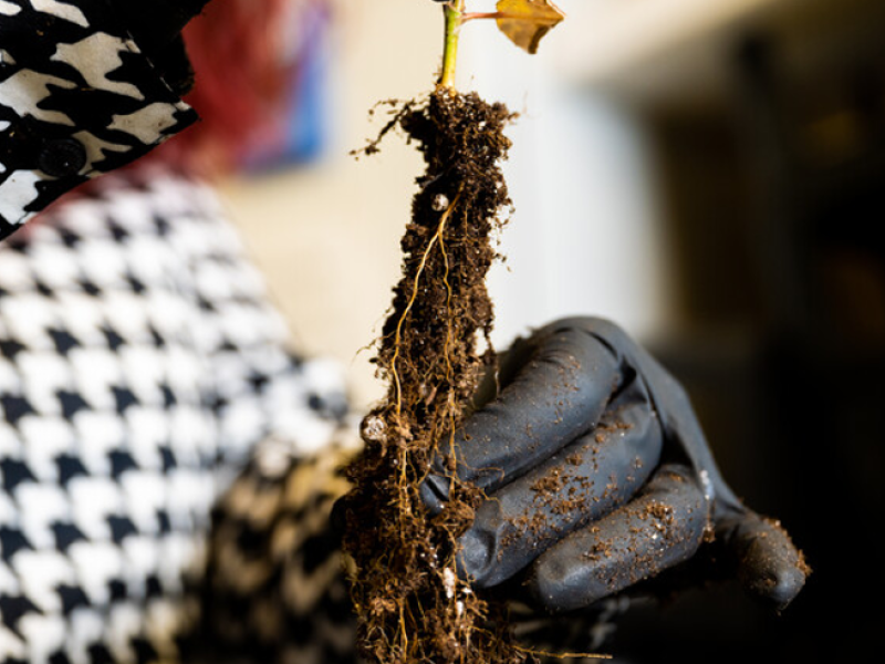
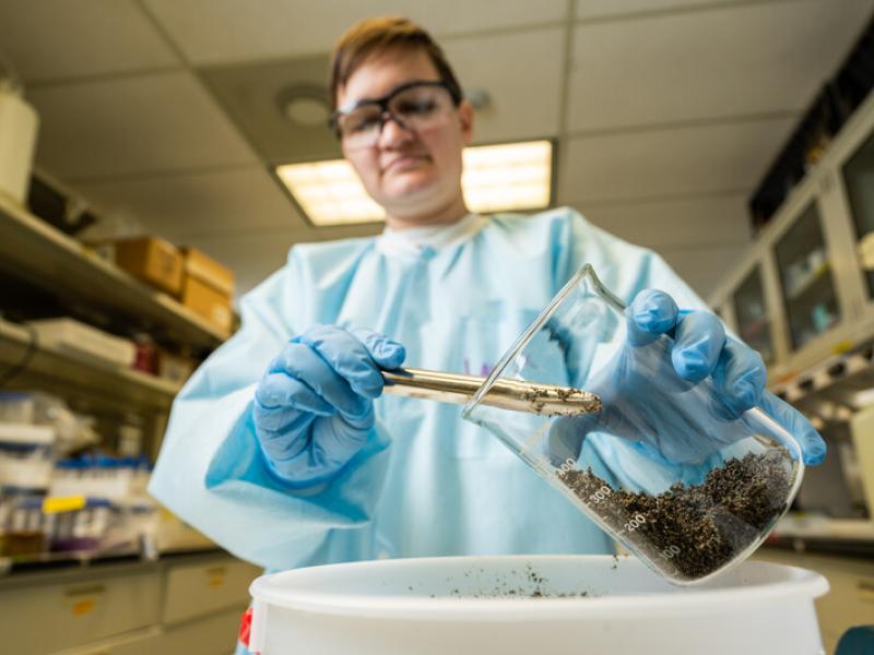
A multi-institutional team of researchers conducted a 13C-labeling greenhouse study using a semi-arid grassland soil, where they tracked the fate of 13C-labeled inputs from living roots and decaying roots from annual grass Avena barbata.
A team of researchers from Pacific Northwest National Laboratory and the Environmental Molecular Sciences Laboratory developed a new and flexible software tool called “Advanced Spectra PCA Toolbox.”
Sequencing of microbiome and characterization of metabolome revealed significantly different functions of fine root systems from four temperate tree species in a 26-year-old common garden forest.
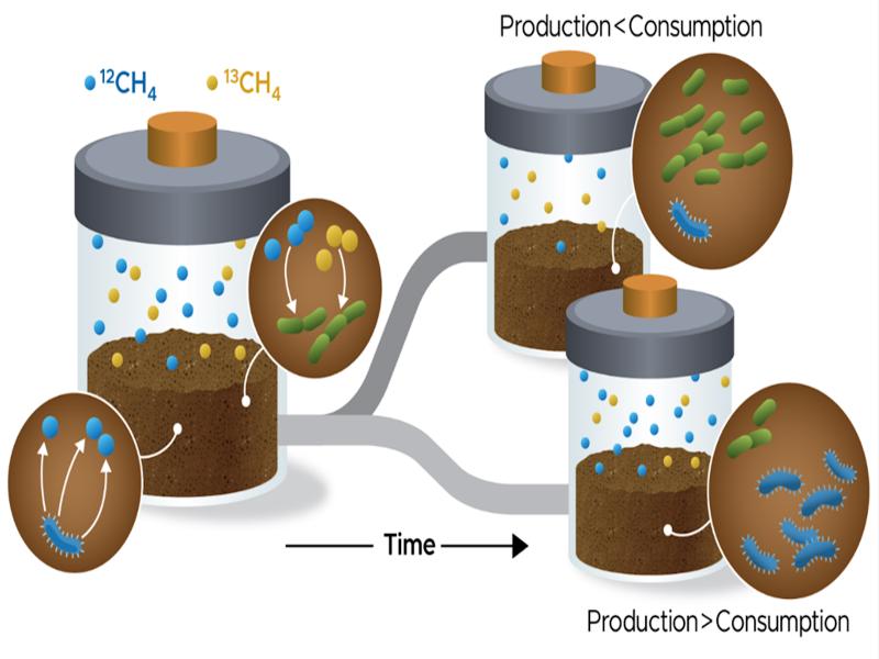
In soil, microbes produce and consume methane. Using a technique called pool dilution, researchers can separate the rate of methane production and consumption from the net rate.
PNNL’s Center for the Remediation of Complex Sites convened attendees from around the world to discuss challenges associated with environmental contamination.
Spatial proteomics enables researchers to link protein measurements to features in the image of a tissue sample, which are lost using standard approaches.