New Imaging Approach Reveals Molecular Changes in Diseased Lung Tissue
Researchers developed a new technique that combined mass spectrometry and label-free optical imaging to map molecular and structural changes in healthy and diseased lung tissue, providing new insight into bronchopulmonary dysplasia

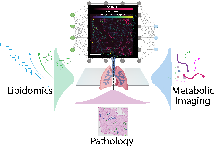
By combining mass spectrometry and label-free optical imaging, researchers developed a new way to study lung disease that reveals both the structure of lung tissue and the molecules within it in greater detail.
(Graphic by artacet | iStock)
The Science
A team of researchers from Pacific Northwest National Laboratory, the University of California San Diego, and the University of Rochester Medical Center developed a new way to study lung tissue by combining several advanced imaging technologies on the same sample. Using a combination of mass spectrometry imaging, which maps hundreds of molecules across tissue, with label-free optical imaging, which reveals tissue structure and metabolism without dyes or stains, they were able to see both the structure of the lung and the specific molecules present down to very small regions (a few cells). Using this approach, they discovered important differences between healthy lungs and those affected by bronchopulmonary dysplasia (BPD), including changes in fats (lipids), energy-related metabolites, and collagen organization. These insights can further reveal how BPD alters the lung at a detailed level and may help guide future therapies.
The Impact
This research provides a new way to study lung disease by allowing scientists to see both the structure of lung tissue and the molecules within it with unprecedented detail. By combining multiple imaging technologies on the same tissue section, the team created a powerful new tool that can reveal hidden biological changes in conditions like BPD. This approach could guide the development of better diagnostics and treatments, and it lays the foundation for future studies that map diseases at the level of individual cells and tissue regions.
Summary
To better understand how bronchopulmonary dysplasia (BPD) alters the lung, a multi-institutional team combined several complementary imaging approaches to examine the same tissue sample. Researchers used high-resolution Fourier transform ion cyclotron resonance mass spectrometry imaging at the Environmental Molecular Sciences Laboratory (EMSL), a Department of Energy Office of Science user facility, to map hundreds of lipid molecules across lung tissue. They paired these measurements with label-free optical imaging from the University of California San Diego to reveal tissue metabolism, protein structures, and collagen organization, then aligned the resulting images into a single, high-resolution view. By bringing together molecular, metabolic, and structural information from the same tissue section, the approach allows researchers to directly connect biological changes with the precise locations where they occur—providing a more complete picture of disease than any single imaging method can achieve alone. This integrated workflow revealed previously difficult-to-observe differences between healthy and diseased lung tissue and provides a foundation for future studies aimed at improving the understanding, diagnosis, and treatment of lung disease.
Contacts
Christopher Anderton
Pacific Northwest National Laboratory | EMSL
Anderton@pnnl.gov
Brittney Gorman
Pacific Northwest National Laboratory | EMSL
Brittney.Gorman@pnnl.gov
Lingyan Shi
University of California San Diego
l2shi@ucsd.edu
Funding
This work was supported by the National Institutes of Health (NIH) National Heart, Lung, and Blood Institute Molecular Atlas of Lung Development Program (LungMAP) Human Tissue Core BioRespository for Investigation of Diseases of the Lung (U01HL122700 and U01HL148861). Additional support was provided by a National Heart, Lung, and Blood Institute grant (U01HL148860) and the NIH Common Fund Human Biomolecular Atlas Program (U54 HL165443-01).
Published: September 25, 2026
B. Gorman; Li, Z.; Deutsch, G.; Huyck, H.L.; Beishembieva, N.; Bhotika, H.; Olson, H.; Villazon, J.; Yu, P.; Pryhuber, G.S.; Clair, G.; Shi, L.; and Anderton, C.R. “Multiscale Metabolic Mapping of Lung Tissue via Coregistered Mass Spectrometry and Nonlinear Optical Imaging.” Science Advances 12, eaec3544 (2026). [DOI: 10.1126/sciadv.aec3544]