Researchers at PNNL share a research- and practitioner-informed approach to assess the threat landscape, elicit and integrate feedback into solutions, and ultimately share outcomes with the emergency response and public safety community.
Two new publications provide emergency response agencies with critical insights into commercially available unmanned ground vehicles used for hazardous materials response.
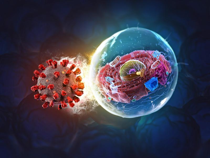
Continued studies will deepen scientists’ understanding of virus-host interactions at the molecular level and also pave the way for developing better drugs to fight emerging viruses.
A team from PNNL contributed several articles to the Domestic Preparedness Journal showcasing recent efforts to explore the emergency management and artificial intelligence research and development landscape.
Researchers developed a robust, cost-effective, and easy-to-use cap-based technique for spatial proteome mapping, addressing the lack of accessible proteomics technologies for studying tissue heterogeneity and microenvironments.
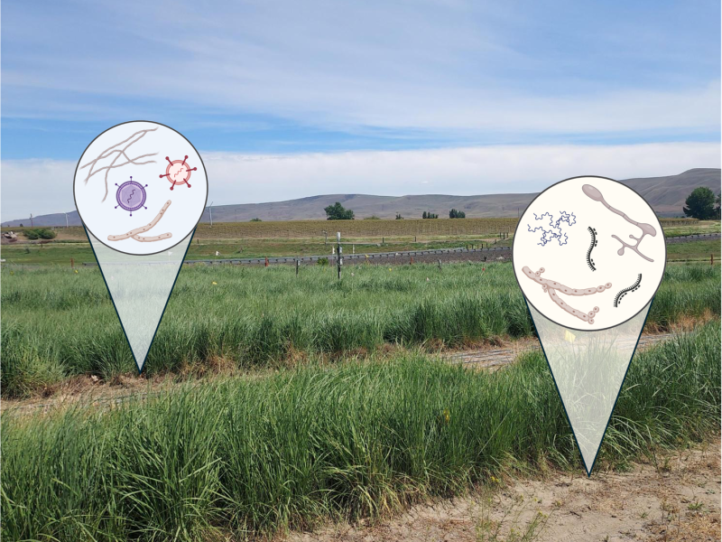
Despite the widespread presence of RNA viruses in soils, little is known about the relative contributions and interactions of biological and environmental factors shaping the composition of soil RNA viral communities.
Early life exposure to polycyclic aromatic hydrocarbons (PAHs), found in smoke, has been linked to developmental problems. To study the impacts of these pollutants, PAH metabolism in infants and adults were compared.
A team of researchers from Pacific Northwest National Laboratory and the Environmental Molecular Sciences Laboratory developed a new and flexible software tool called “Advanced Spectra PCA Toolbox.”
Discovering and measuring the spatial organization of proteins within cells allows scientists to map complex proteoforms across tissues with near-cellular resolution.
PNNL advisors joined a panel of Washington State emergency management personnel to discuss how partnerships with national laboratories are enabling science and technology solutions.