New Views of Protein Biology: Visualizing Proteoforms Directly from Tissues
Discovering and measuring the spatial organization of proteins within cells allows scientists to map complex proteoforms across tissues with near-cellular resolution

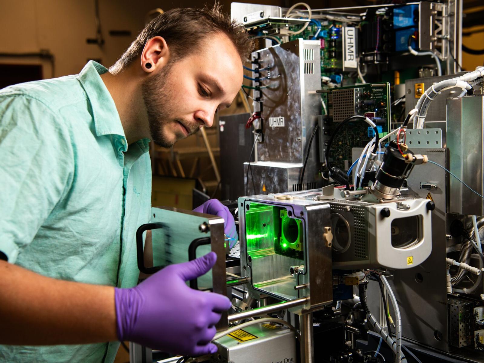
Mass spectrometry imaging enables high-throughput multiplexed proteoform mapping directly from tissue.
Andrea Starr for Pacific Northwest National Laboratory
The Science
Proteoforms, or multiple forms of the same protein, can result from genetic variation and different regulatory mechanisms (e.g., posttranslational modifications). Unique gene products, proteoforms, help us understand protein function and therefore can be used as biomarkers of a cell’s state (i.e., diseased, healthy). In this study, researchers developed an integrated workflow to detect proteoforms and reveal their spatial distributions. This mapping of proteoforms across tissues could help us discover additional biomarkers and enable precision medicine for disease treatment.
The Impact
While there are roughly 20,000 proteins in the human body, there are an estimated tens of millions of unique proteoforms. Detection of proteoforms directly from tissues is challenging, and despite the potential to better inform biological outcomes via biomarker discovery and precision medicine, we lack routine methodology for sensitive high-throughput proteoform mapping. In this study’s scope, researchers demonstrated proteoform detection down to 15 µm spatial resolution, meaning they can directly measure previously unseen chemical diversity among cells within functional tissue units. Having the capability to profile small, biologically active proteoforms will enable high-impact biomedical and environmental (e.g., structures of biofilm, plant cell differentiation, etc.) discoveries.
Summary
Through the Human BioMolecular Atlas Program (HuBMAP), a National Institutes of Health Common Fund consortium focused on developing an open global platform for mapping normal states of healthy cells in the human body, researchers in this study highlighted the importance of proteoforms. The unique spatial top-down proteomics pipeline they developed provides new perspectives into the fundamental mechanisms of cellular function. The core of the pipeline is broad field-of-view imagery produced through a technique called matrix-assisted laser desorption/ionization, where proteoforms are directly ionized from thin tissue sections and can be visualized at cellular resolution. The researchers also used a laser to microdissect portions of thinly sliced tissue using a method called laser capture microdissection. In tandem with nanodroplet or microdroplet processing in one pot for trace samples (nanoPOTS/microPOTS) technology, also developed at Pacific Northwest National Laboratory (PNNL), the researchers captured snapshots of the diverse proteome from entire functional tissue units or small regions of interest with the highest levels of sensitivity. This detailed information about where and how proteoforms were localized within tissue gives clues about a protein’s response to disease or environmental cues.
Contact
Ljiljana Paša-Tolić, ljiljana.pasatolic@pnnl.gov, PNNL
Kevin J. Zemaitis, kevin.zemaitis@pnnl.gov, PNNL
James M. Fulcher, james.fulcher@pnnl.gov, PNNL
Funding
Research and development was funded by the National Institutes of Health Common Fund, HuBMAP grant UG3CA256959, and was performed at the Environmental Molecular Sciences Laboratory (EMSL), a Department of Energy, Office of Science user facility sponsored by the Biological and Environmental Research program under Contract No. DE-AC05-76RL01830, on project award doi.org/10.46936/staf.proj.2020.51770/60000309.
Additional instrument development was funded by the Science and Technology intramural program at EMSL on project award doi.org/10.46936/intm.proj.2019.51159/60000152.
Published: June 7, 2024
Zemaitis, K.J., Fulcher, J.M., Kumar, R. et al. Spatial top-down proteomics for the functional characterization of human kidney. Clin Proteom 22, 9 (2025). https://doi.org/10.1186/s12014-025-09531-x