Technology Overview
Structured laser light enhances lateral resolution and suppresses out-of-focus blur
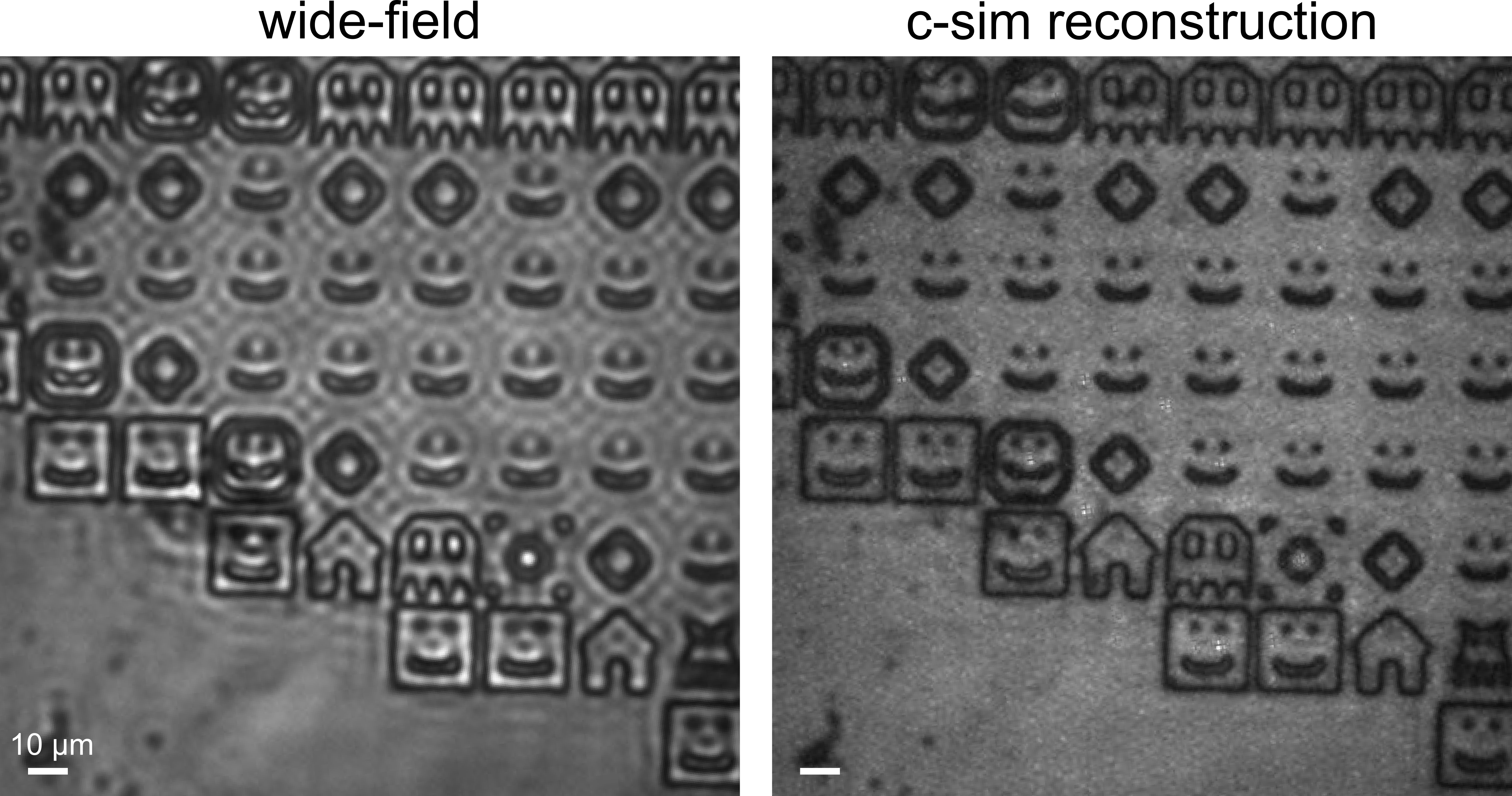
Coherent structured illumination microscopy (C‑SIM) is a microscopy technique that uses patterned laser light to create sharper, clearer images of unlabeled samples such as cells, particles, or materials. By shining multiple angled light beams onto a sample and by computationally combining the resulting images, it can reveal fine details that normal microscopes blur out. Pacific Northwest National Laboratory (PNNL) has developed a new version of C-SIM that improves both image sharpness and the ability to “see through” out‑of‑focus layers, making structures inside thick or highly scattering samples much easier to distinguish.
Improving optical sectioning
In optical microscopy, coherent laser light is often used to access new sample information, but it has difficulty separating information from different sample planes. Optical sectioning is a microscopy approach to circumvent this that creates clear images from a single, thin “slice” of a sample by suppressing light that comes from out‑of‑focus depths. Instead of physically cutting a specimen, the microscope uses optical patterns or filtering techniques to reject blurry background light, allowing only the in‑focus plane to contribute to the image. In C‑SIM, this is achieved by using structured illumination, which more effectively removes out‑of‑focus contributions, producing sharper, cleaner images.
Advantages
The key benefits of C‑SIM include the following:
- Sharper images beyond the normal resolution limit
- C‑SIM provides up to a 2× improvement in lateral resolution, allowing much finer structural details to be seen compared to those obtained with standard wide‑field imaging.
- Works on nonfluorescent, light‑scattering samples
- Unlike traditional SIM (which typically requires fluorescence), C‑SIM works with label‑free samples, including biological cells and materials that scatter light.
- Enhanced optical sectioning (especially in five‑beam mode)
- The five‑beam configuration adds an on‑axis illumination component that significantly reduces out‑of‑focus blur and coherent defocus artifacts, yielding clearer images in thick or complex samples.
- Cleaner images with less ringing and speckle
- C‑SIM suppresses coherent artifacts such as ringing, speckle, and grainy backgrounds, leading to more natural‑looking images. The five‑beam version improves this even further.
- Fast, intensity‑only imaging (no complex interferometry)
- Because C‑SIM detects intensity images only, it avoids the complexity of measuring optical phase fields, simplifying the setup and enabling fast 2D imaging.
- Useful for live or delicate samples
- Demonstrations on live microalgae show that C‑SIM can improve contrast and detail without labeling, making it suitable for live‑cell, label‑free imaging.
INDUSTRY APPLICATIONS
C‑SIM can benefit industries that rely on high‑clarity, label‑free imaging of complex or scattering materials. In biotechnology and the life sciences, it can improve the visualization of cells, tissues, and microorganisms without fluorescent tags. In semiconductor and advanced materials manufacturing, its enhanced resolution and reduced imaging artifacts can help detect surface defects or variations. Environmental science, energy research, and clean‑tech applications can use C‑SIM to study heterogeneous materials with greater accuracy. Its ability to provide clearer, faster, nondestructive imaging also makes it attractive for industrial research and development (R&D) labs and quality‑assurance workflows.