Radiological Microscopy Suite
Instrumentation magnifies PNNL's research
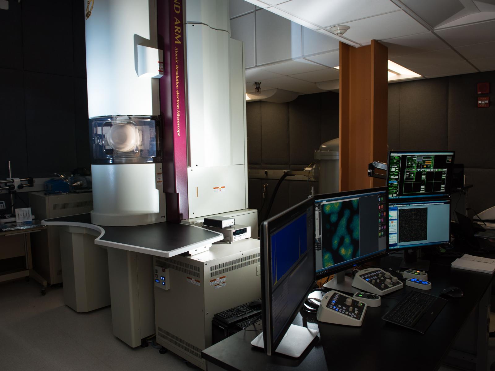
The Radiological Microscopy Suite at PNNL is home to a JEOL GrandARM-300F STEM. This unique instrument allows unprecedented atomic-scale characterization of nuclear materials, structural alloys, and functional systems in dynamic conditions.
(Photo by Andrea Starr | Pacific Northwest National Laboratory)
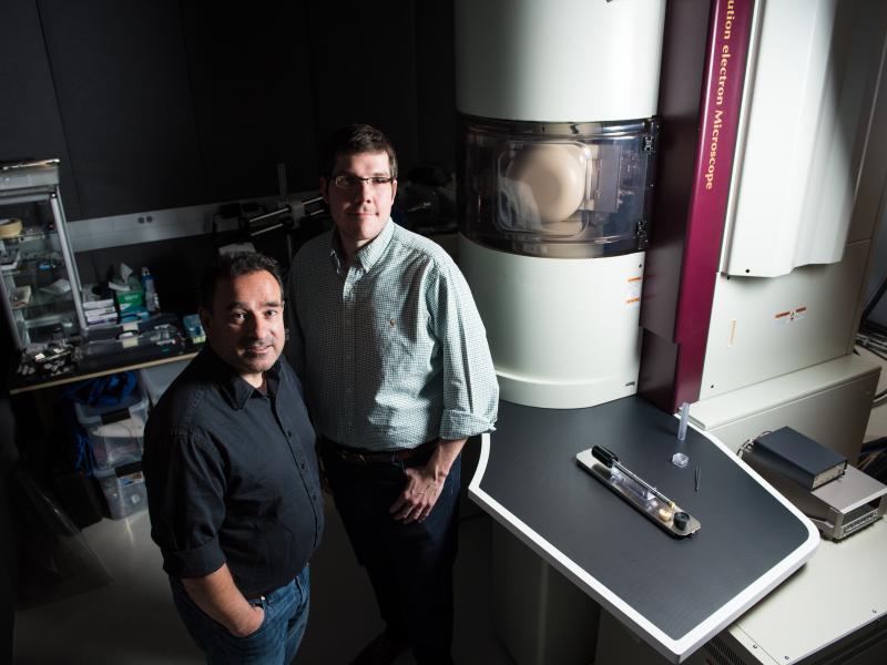
PNNL scientists Edgar Buck and Steven Spurgeon in the Radiological Microscopy Suite, home to a JEOL GrandARM-300F STEM. This unique instrument allows unprecedented atomic-scale characterization of nuclear materials, structural alloys, and functional systems in dynamic conditions.
(Andrea Starr | Pacific Northwest National Laboratory)
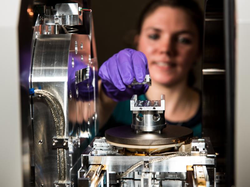
Post doctorate student Bethany Mathews uses the Thermo Fisher Helios 660 Dual Beam FIB-SEM for preparation and analysis of radioactive and non-radioactive samples, including metals, oxides, and geologic materials.
(Andrea Turner | Pacific Northwest National Laboratory)
Post doctorate student Bethany Mathews uses the Thermo Fisher Helios 660 Dual Beam FIB-SEM for preparation and analysis of radioactive and non-radioactive samples, including metals, oxides, and geologic materials.
(Andrea Starr | Pacific Northwest National Laboratory)