FEI Helios 660 Focused Ion Beam-Scanning Electron Microscope
Located in RPL

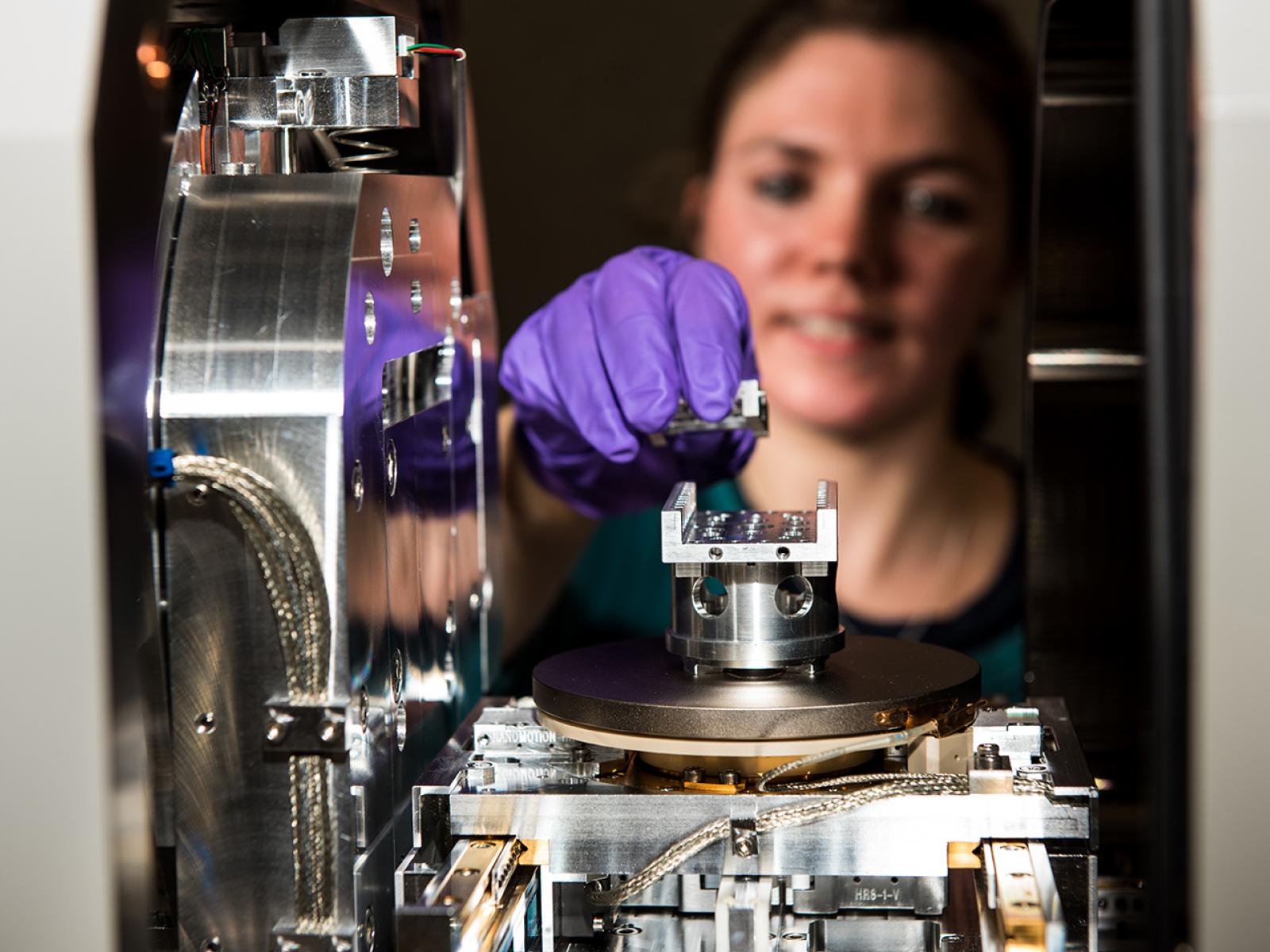
Post doctorate student Bethany Mathews uses the Thermo Fisher Helios 660 Dual Beam Focus Ion Beam Scanning Electron Microscope (FIB-SEM) for preparation and analysis of radioactive and non-radioactive samples, including metals, oxides, and geologic materials.
The “dual” part of the microscope is what makes it special. Researchers can prepare samples and analyze them while also using one of the beams to remove site-specific material from the sample.
Pacific Northwest National Laboratory | Andrea Starr
Mission
The FEI Helios 660 Focus Ion Beam Scanning Electron Microscope (FIB-SEM) is a workhorse for both radiological and non-radiological material sample preparation and analysis in energy, environment, and national security research.
It’s ideal for micron to nano scale imaging, chemical analysis, and sample preparation for transmission electron microscope (TEM) lamella, atom probe tomography (APT) and secondary ion mass spectrometry (SIMS) analysis, including metals, powders, and thin films. High-resolution surface composition mapping enables researchers to understand minute details of a material and its degradation over time in certain environments.
Features
- Gallium Ion source 30 keV
- Schottky Electron source
- EasyLift for extracting FIB-TEM, APT and SIMS samples
- Low keV retractable backscatter detector
- Retractable scanning/transmission electron microscopy detector
- EDAX energy-dispersive detector and electron backscatter diffraction detector
- Bruker Micro CT scanner
- Transfer arm for vacuum transfer
Related publications:
Spurgeon SR, M Sassi, ES Ilton, EC Buck. 2019. "First atomic-scale actinide spectroscopy sheds light on key material: . Nanoscale Oxygen Defect Gradients in UO 2+ x Surfaces." Proceedings of the National Academy of Sciences of the USA, Vol. 116, Issue 35, 17181-17186.