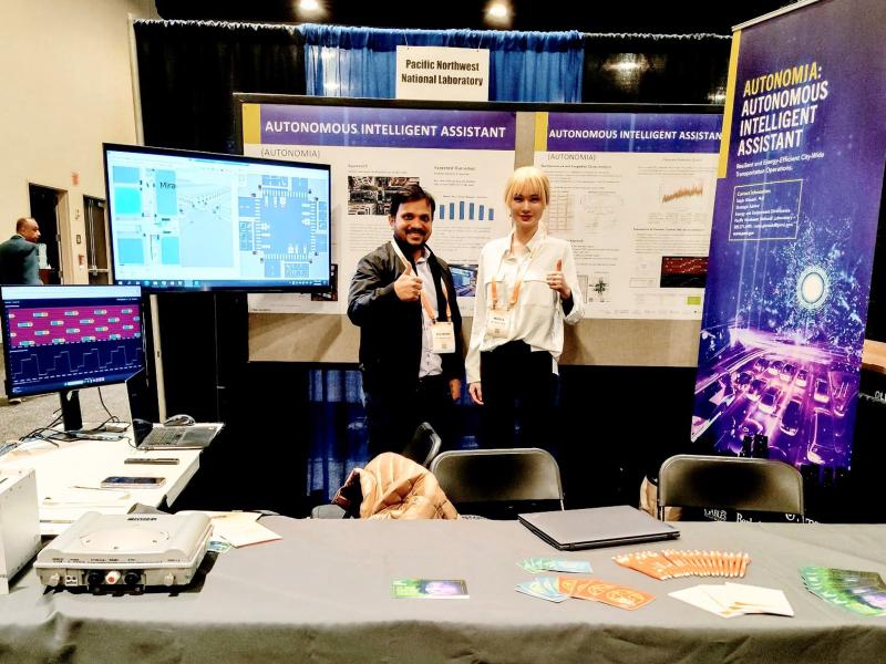
The ARPA-E Energy Innovation Summit brings together researchers, industry leaders, entrepreneurs, and investors to showcase the latest technologies shaping tomorrow’s energy landscape. This year, eight projects led by PNNL were featured.
Researchers developed a robust, cost-effective, and easy-to-use cap-based technique for spatial proteome mapping, addressing the lack of accessible proteomics technologies for studying tissue heterogeneity and microenvironments.
PNNL researchers are exploring the kinds of flicker waveforms that the eye and brain can detect, seeking to understand the different visual and non-visual effects that result.
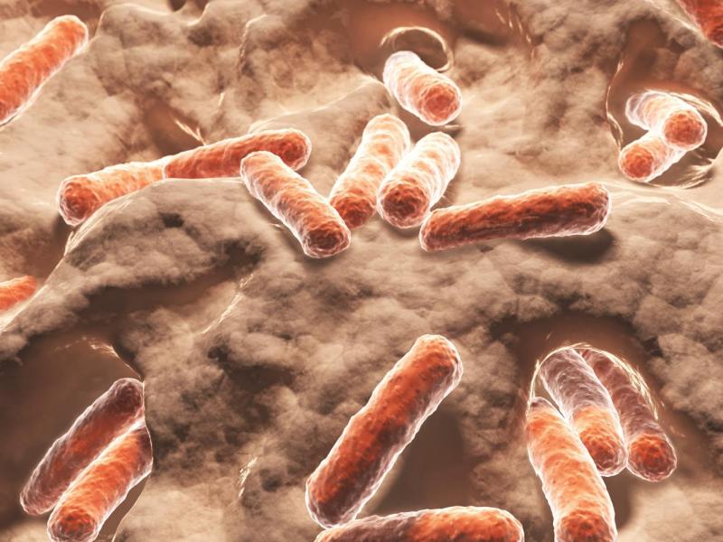
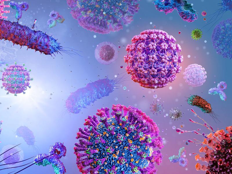
Despite the widespread presence of RNA viruses in soils, little is known about the relative contributions and interactions of biological and environmental factors shaping the composition of soil RNA viral communities.
A compilation of soil viral genomes provides a comprehensive description of the soil virosphere, its potential to impact global biogeochemistry, and an open database for future investigations of soil viral ecology.
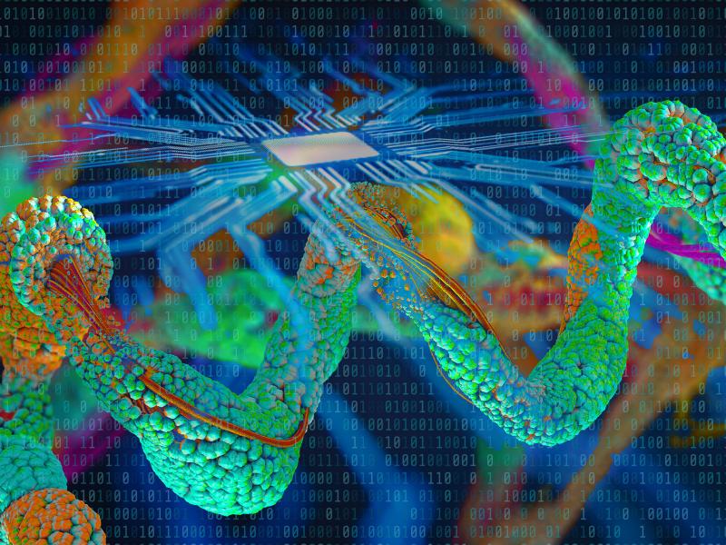
A team of researchers from Pacific Northwest National Laboratory and the Environmental Molecular Sciences Laboratory developed a new and flexible software tool called “Advanced Spectra PCA Toolbox.”
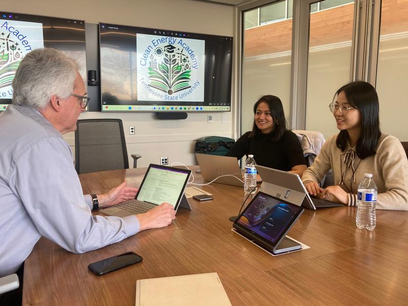
Tennessee State University received Department of Energy funding to establish an academy focused on preparing students and professionals to work in an emerging field: clean energy systems. PNNL is helping with that effort and others.
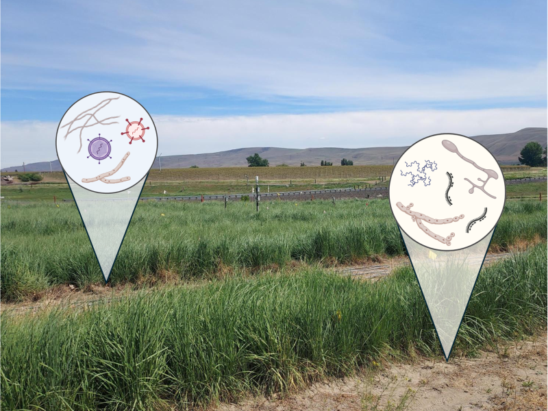
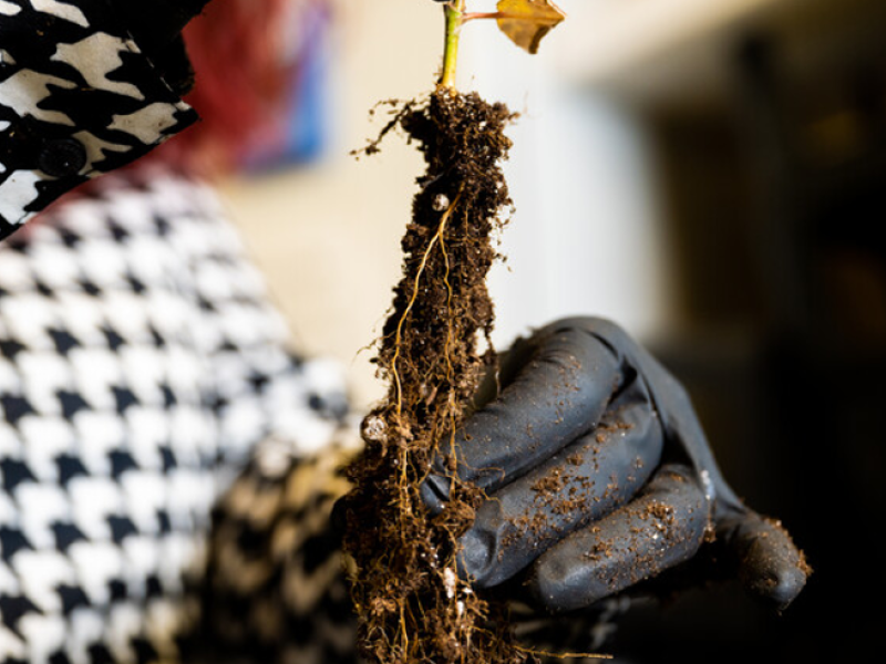
Sequencing of microbiome and characterization of metabolome revealed significantly different functions of fine root systems from four temperate tree species in a 26-year-old common garden forest.
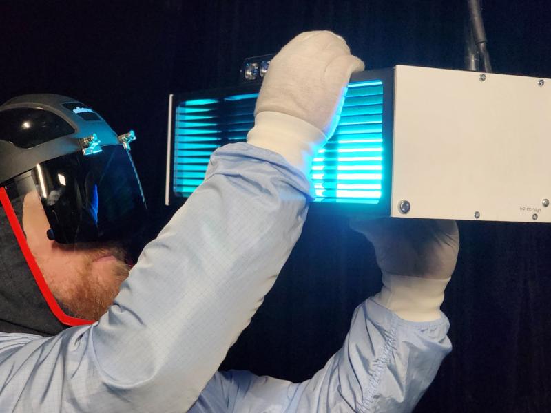
GUV can reduce transmission of airborne disease while reducing energy use and carbon emissions. But fulfilling that promise depends on having accurate and verifiable performance data.