Photoemission Electron Microscope
Located in EMSL | Stewarded by Alan Joly, Chemical Physics and Analysis Experimental Team
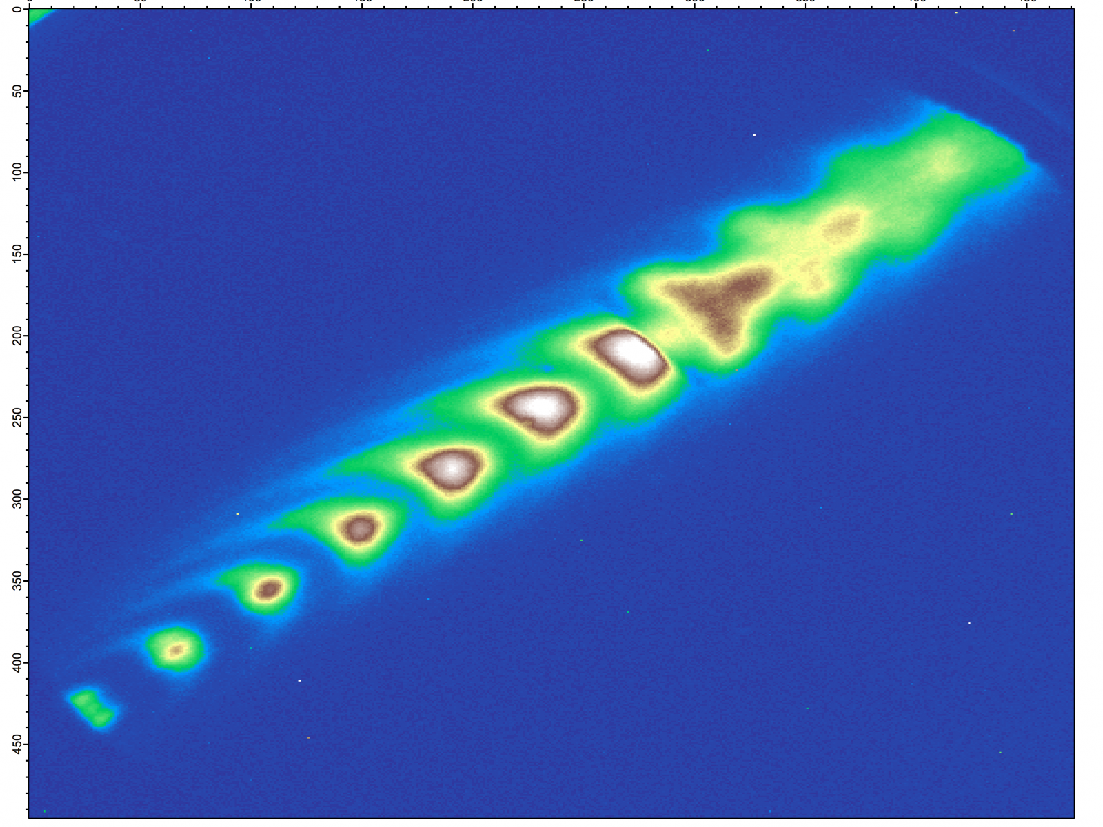
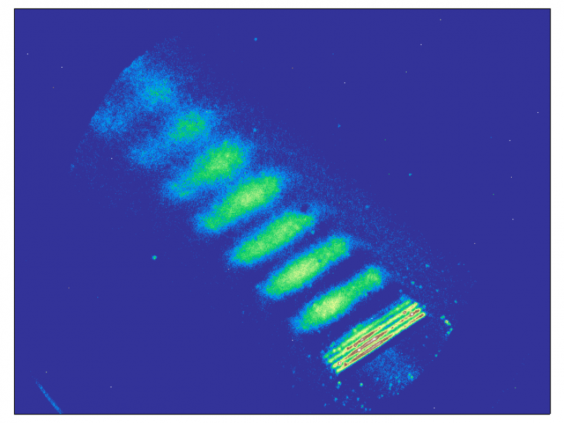
Photoemission electron microscope (PEEM) images depicting surface plasmon propagation on thin silver surfaces.
Alan Joly
Mission
The photo-emission electron microscope (PEEM) is capable of imaging nanoscale surface structures (~20 nm) via electron emission induced by ultraviolet and laser light sources. The PEEM is applied to surface science studies of individual nanostructures and tailored plasmonic constructs. Experiments can be performed with either static or dynamic light sources. In particular, femtosecond laser excitation allows pump-probe studies of surface plasmon propagation revealing the underlying physics required to turn nanoscale structures into miniaturized nanophotonic circuits.
Features
- Spatial resolution up to 20 nm
- Continuous to femtosecond temporal resolution
- Still to continuous motion image modes
- Metal and semiconductor sample types
- 150-2 micrometer field of view
- Base pressure of 1×10⁻¹⁰ Torr
- Sample temperature of liquid nitrogen to 900°C