May 30, 2019
News Release

New Technique Sees Bacterial Growth Quicker

Approach has implications for food safety, health and research

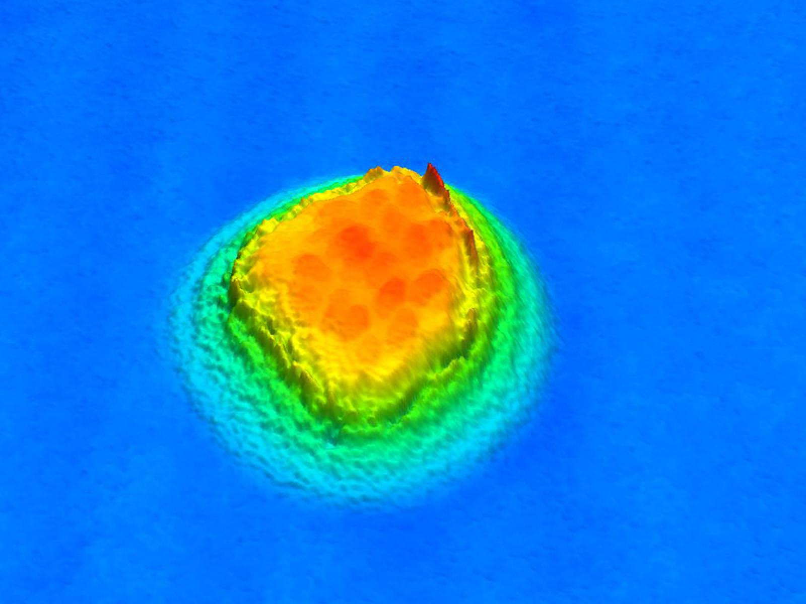
Bacteria colony

The new technique lets researchers have a close-up view of bacterial colonies like this one. The cells are about 100 times smaller than a human hair.

RICHLAND, Wash. - Whether a clinical lab technician is helping a doctor know if a patient has a bacterial infection or a researcher is studying bacteria looking for a new treatment, it is still common to grow cells into bacterial colonies on a classic Petri dish with its nutrient medium to count how many cells are in a sample. This tried-and-true method takes time and resources for cells to replicate enough that they form a colony that can be seen and counted with the naked eye. A similar process is employed in food processing plants. In many cases, time is of the essence.

Researchers at PNNL have developed a patent-pending technique for a faster and improved way to count bacteria and measure their growth, without killing the sample, by using a white light interferometer. 

WLIs are microscopes that split a beam of light and then look for characteristic wavy patterns that appear in images as the two halves of the light interfere with each other when recombined. The interference patterns translate into a measurement of different heights or textures on the surface of what’s being viewed and can track it as it changes.

Using this previously unexplored approach, researchers were able to quickly see, in great detail, barely perceptible changes in heights down to three nanometers – more than 10,000 times smaller than the diameter of a human hair – in the topography of a sample. Being able to see very detailed height changes is important for bacterial colonies which, the team observed, grow slowly upward as well as out.

“What is unique and what is really powerful about this technique is that it gives you unprecedented resolution in the vertical direction,” said PNNL materials scientist Curtis Larimer. “That means you can look at a wide area and still see all these really incredible fine details.”

This fine detail allows investigators to take a single image and determine if bacterial colonies are present. The technique is much faster than traditional approaches. They visualized and counted bacteria on a Petri dish within two to six hours of placing the sample on the dish, compared to the typical incubation time of one to three days or longer, depending on the sample species in question. PNNL researchers reported their findings in Scientific Reports, a journal published by Nature.

A Broad View of Bacterial Colonies  

With regular microscopes, to look at finer details, you have to zoom in, which vastly shrinks the field of view, cutting off valuable information in the rest of a sample. And typically, you have to zoom in pretty far before you can start to see very small bacteria and colonies that are just beginning to form. With WLI you get the same detailed vertical resolution in a zoomed-out view as you get when zoomed in. And the vertical resolution is about 100 times better than one can see with a standard light microscope.

Researchers at PNNL have developed a patent-pending technique for a faster and improved way to count bacteria and measure their growth, without killing the sample, by using a white light interferometer.

WLIs are not new. In fact, the concept dates back to Isaac Newton’s study of ring patterns in optical glasses in the 18th century. They can easily measure how rough or smooth something is and are used commercially for things like quality control on optical lenses and silicon chips. But they are not used on biological systems because WLI was designed to work on hard, dry, reflective surfaces. However, the PNNL team perfected a technique for using it on wet, rough samples, which are needed to observe bacterial growth.

Tests in the lab created 3-D images of two common classes of bacteria growing on Petri dishes: a Gram-negative species (Pseudomonas fluorescens) and a Gram-positive species (Bacillus thuringiensis). Researchers were able to accurately count colony-forming units while also accurately quantifying growth rates and other structural features.

The team developed software that works behind the scenes to analyze the image, remove the noise in the data, while preserving the signal from the bacteria and reconstruct the numbers into an early indicator, 3-D picture that should be useful for doctors or researchers.

“We are not clinicians but we envision, with this new approach to measuring bacterial counts, a patient could provide a sample and within two to six hours, the doctors would know in fact that it is a bacterial infection and what types of treatment would work best,” said Larimer. “Rapid information about bacterial growth could be part of the key to thwarting antibiotic resistant drugs.”

Used in a food processing plant, the team believes that food-borne bacteria could be identified and stopped before a contaminated product hits the store shelves, keeping consumers safe and avoiding massive product recalls for the company.

Building on Biofilm Research

The WLI technique can count bacteria in a sample but isn’t able to determine specific strains. However, another benefit is that it does not destroy the sample, which could be subjected to other techniques for identification. And, it may be possible to automate further analysis of the image that would point to the specific type of bacteria.

Researchers received funding from PNNL’s Microbiomes in Transition Initiative to build on earlier work on biofilms. The same research team previously developed a material designed to prevent the formation of biofilms and biofouling on things like ships’ hulls and water intake systems. In that process, they needed to measure biofilms and pulled together an interdisciplinary team of materials scientists, biologists and computational experts who decided to develop a way to use WLI for those measurements. The diverse team saw a need to further develop the technique, which has a patent pending for bacterial growth for health, research and food safety applications.

The PNNL research team includes Larimer, Jonathan Suter, and corresponding author R. Shane Addleman, as well as former PNNL researchers Michelle Brann, Joshua Powell and Matthew Marshall.

Information on licensing of this technology is available from commercialization manager Jennifer Lee.

About PNNL

Pacific Northwest National Laboratory draws on signature capabilities in chemistry, earth sciences, and data analytics to advance scientific discovery and create solutions to the nation's toughest challenges in energy resiliency and national security. Founded in 1965, PNNL is operated by Battelle for the U.S. Department of Energy's Office of Science. DOE's Office of Science is the single largest supporter of basic research in the physical sciences in the United States and is working to address some of the most pressing challenges of our time. For more information, visit PNNL's News Center. Follow us on FacebookInstagramLinkedIn and Twitter.

###

About PNNL

Pacific Northwest National Laboratory draws on its distinguishing strengths in chemistry, Earth sciences, biology and data science to advance scientific knowledge and address challenges in energy resiliency and national security. Founded in 1965, PNNL is operated by Battelle and supported by the Office of Science of the U.S. Department of Energy. The Office of Science is the single largest supporter of basic research in the physical sciences in the United States and is working to address some of the most pressing challenges of our time. For more information, visit the DOE Office of Science website. For more information on PNNL, visit PNNL's News Center. Follow us on Twitter, Facebook, LinkedIn and Instagram.
Published: May 30, 2019