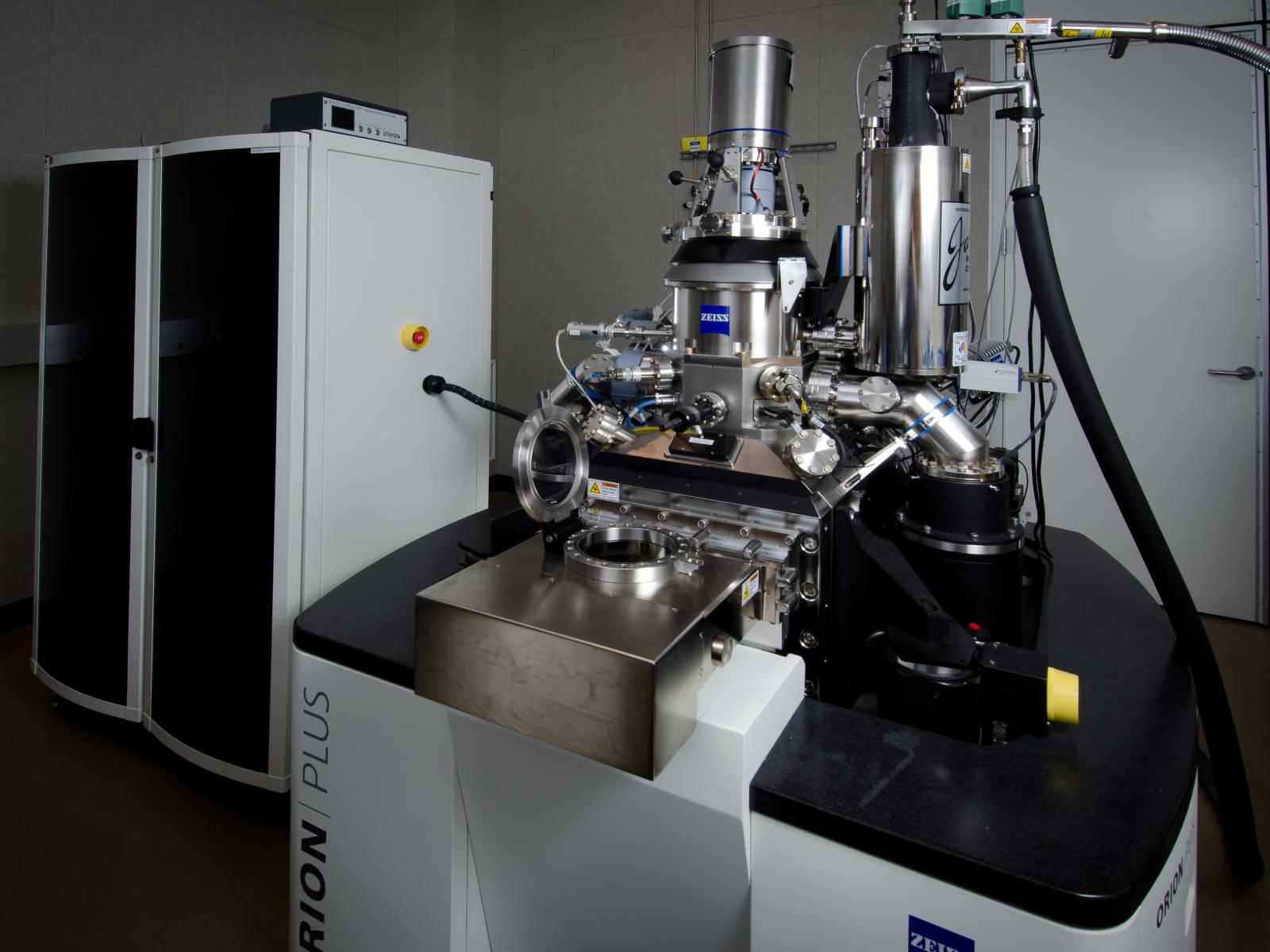
Mission
The Helium Ion Microscope (HIM) advances biological, geochemical, biogeochemical, and surface/interface studies using its combined surface sensitivity and high spatial resolution. The high-resolution (0.35 nm) and outstanding depth of field (few microns) that can be achieved for the imaging of uncoated biological and mineral material makes the HIM especially suitable in the biological and environmental research domain. The HIM is equipped with a Rutherford backscattering spectrometry and imaging capability that identifies atomic elements and provides Z contrast images.
Features
- Ultra-high resolution: Reveals fine structure and surface sensitive details that allows for the chemical visualization of nanostructures in biological, mineral, and environmental samples, including those with low-Z elements. HIM has the best secondary electron imaging resolution ~ 0.35 nm obtained in any microscope.
- High surface sensitivity: The large signal to noise ratio gives enhanced surface details and high contrast between high and low Z elements.
- Large depth of field enables visualization of three-dimensional structures providing detailed spatial relationships between various components (e.g., microbes, plant root, mineral, organic matrix) of a sample.
- Minimum beam damage: Small interaction volume between the helium beam and the sample results in lesser energy transfer (damage) to the surface layers. Hence, beam sensitive samples, such as polymers and organic materials, that are readily damaged in scanning electron microscope (SEM) can be imaged with HIM with minimal beam effects.
- Conductive coatings are not necessary to image insulating samples. HIM uses an electron flood gun to neutralize the charge buildup at the surfaces for insulating samples. Because of this capability, highly charging samples like biological and soil organic matter/minerals samples (which can be problematic to image in a scanning electron microscope) can be analyzed without coatings to capture the true surfaces and interfaces.
- Backscattering ion imaging: Since backscattering yield is proportional to Z2, it is possible to get Z contrast images using this mode. In addition, since ion beams can penetrate and there is backscatter from regions below the sample surface, it is possible to get some depth information (like buried interfaces and features).
- Rutherford backscattering spectrometry enables the identification of elements and determines material composition using a sub-nanometer He+ ion probe.
- Ultra-high-resolution lithography: Small probe size and limited interaction volume offers the possibility of patterning small features (nm) with high profile (microns) fidelity using HIM.
- Because of a small beam size (0.35 nm), ion beams from HIM are ideal to perform site specific radiation studies on biological and radiation resistance materials.