CDI Capability: Micro-CT Imaging
Enhancing PNNL’s Micro-CT Imaging Capabilities
A new, micro x-ray computed tomography (micro-XCT or micro-CT) instrument with an increased spatial resolution has been a key need to round out PNNL’s suite of characterization capabilities. The primary driver for this new system is the need to visualize fine structures and enable research at an intermediate scale between the current Nikon XCT and what’s available through beamlines or the new Sigray nano-CT. The system is scheduled for install in May 2020 and will be fitted with a high-resolution LaB6 source, in addition to the standard Tungsten source.

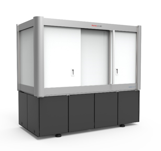
New micro-CT Instrument: Thermo Fisher Heliscan MicroCT Mark II
The HeliScan microCT is a versatile, micro-computed tomography (micro-CT) imaging system that produces geometrically accurate 3D images of sample materials of any type and shape using conventional circular as well as patented helical trajectories. The HeliScan technology enables continuous scanning of tall samples, thus avoiding artifacts commonly associated with stitching. It also produces high-quality images over the entire volume of sample imaged for quantitative image analysis. Thermo Fisher Scientific’s scanning technology uses wide X-ray cone angles and a high-flux imaging environment for fast data recording low noise images that accurately represent a sample’s microstructure without distortion and are ready for segmentation and analysis.
Main Specifications:
- Voltage: 20-160 kV, Power: 16 W, Three optimized focus modes: Small, Medium and Large
- Maximum sample diameter: 24 cm
- Spatial resolution: 800 nm Tungsten target, 400 nm LaB6 target
- Nominal resolution (Voxel size at the highest possible magnification): 170 nm
The HeliScan microCT features a water-cooled X-ray source capable of producing an X-ray beam with uniform flux over an extended period. The instrument also maintains X-ray focus during the entire duration of imaging. A key advantage of this design is that it facilitates the imaging of tall samples and produces sharper images with uniform intensity throughout the image volume. The source is integrated with a nitrogen venting line which allows for contaminant-free working conditions for easy filament exchange and which minimizes downtime. The iterative reconstruction technology provides the critical image resolution and high signal-to-noise acquisition needed for targeted signature analysis and high-confidence measurement.
Supporting the need for high-fidelity measurements and visualization, the new CT lab will have a dedicated workstation powered by Avizo software. Fast reconstruction needs will be supported by a direct link to the GPU resources within the Marianas cluster.